Case 1 Bone mass on Right Shoulder
14 year old student presented ith a painful lump on the right shoulder. X-rays showed the bony mass which looked benign.
Due to its size and position, removal was anticipated to be difficult and the proximity to several nerves increased the risk of complications.
A plain CT scan was obtained and a 3D model created using TLA software. Because this could be easily and quite conveniently demonstrated to the patient and his parents in the normal consulting room, they could understand both the process to remove the tumour, plus the risks involved. As the muscles could also be visualised, the actual surgical approach could be planned in 3D.
 The tumour was succesfully removed and histology confirm the benign nature. That the patient could appreciate the extent of bone removal and the risk of postoperative fracture ensured compliance during the recovery period.
Case 2 Ectopic bone around Right Elbow
27 year old male who was involved in a crushing injury to the right arm. There was extensive soft tissue damage anterior to the elbow and the vascular injury was addressed with a vein graft to the brachial artery. The skin defect was covered with a free lattisimus dorsi muscle free flap, which was anastomosed to the vein graft. Post operative progress was complicated by ectopic bone formation around the elbow, blocking motion.
Removing the abnormal bone was going to be difficult due to involvement of the the vein graft and associated free flap. By obtaining a CT arteriogram using a IV bolus of contrast in the opposite arm, the vessels could be visualised and, using TLA software, a 3D image was created from the plain CT data. This image could then be accessed in the Operating Theatre by the surgeon in real time to visualiise the bone and vasular relationship as desired. The exact position of the free flap vessels could be determined by direct measurement, and the strategic bone connections identified.
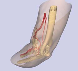
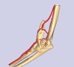
The ectopic bone was removed succesfully without damaging any of the vascular structures, and the patient regained better motion. Further recovery of hand function is anticipated.
Case 3 Pelvic Fracture
37 year old motor cyclist struck by a car. Severe injuries to left hemi pelvis and on plain x-rays the optimum approach was difficult to define. Even on plain CT scan the fracture conformation was not well understood.
Using TLA software a 3D image of the pelvic fracture was created from the plain CT data and this allowed the surgeon to plan crarefully the approach, anticipate the fixation technique required and then expain and show the patient what was involved.
The fracture was successfully reduced and stabilised. The surgeon was able to approach the fracture with much greater confidence having preplanned the operation in 3D pre-operatively.
Case 1 Bone mass on Right Shoulder
14 year old student presented ith a painful lump on the right shoulder. X-rays showed the bony mass which looked benign.
Due to its size and position, removal was anticipated to be difficult and the proximity to several nerves increased the risk of complications.
A plain CT scan was obtained and a 3D model created using TLA software. Because this could be easily and quite conveniently demonstrated to the patient and his parents in the normal consulting room, they could understand both the process to remove the tumour, plus the risks involved. As the muscles could also be visualised, the actual surgical approach could be planned in 3D.
Case 2 Ectopic bone around Right Elbow
27 year old male who was involved in a crushing injury to the right arm. There was extensive soft tissue damage anterior to the elbow and the vascular injury was addressed with a vein graft to the brachial artery. The skin defect was covered with a free lattisimus dorsi muscle free flap, which was anastomosed to the vein graft. Post operative progress was complicated by ectopic bone formation around the elbow, blocking motion.
Removing the abnormal bone was going to be difficult due to involvement of the the vein graft and associated free flap. By obtaining a CT arteriogram using a IV bolus of contrast in the opposite arm, the vessels could be visualised and, using TLA software, a 3D image was created from the plain CT data. This image could then be accessed in the Operating Theatre by the surgeon in real time to visualiise the bone and vasular relationship as desired. The exact position of the free flap vessels could be determined by direct measurement, and the strategic bone connections identified.
The ectopic bone was removed succesfully without damaging any of the vascular structures, and the patient regained better motion. Further recovery of hand function is anticipated.
Case 3 Pelvic Fracture
37 year old motor cyclist struck by a car. Severe injuries to left hemi pelvis and on plain x-rays the optimum approach was difficult to define. Even on plain CT scan the fracture conformation was not well understood.
Using TLA software a 3D image of the pelvic fracture was created from the plain CT data and this allowed the surgeon to plan crarefully the approach, anticipate the fixation technique required and then expain and show the patient what was involved.
The fracture was successfully reduced and stabilised. The surgeon was able to approach the fracture with much greater confidence having preplanned the operation in 3D pre-operatively.
Case 4 3D Spect CT Images
The screen shot images show two levels of activity, on the low res CT GE Hawkeye, showing arthritis in the base of the R 2nd CMC joint, and L distal RU joint.
We have combined the 3D CT data and the bone scan data. There are 3 levels of nuc intensity, with the final darker area showing the area of concern, both in the 2nd metacarpal base, and the dru joint, although the second area did not have much localised symptoms.
The specific localisation in 3D was very helpful as the main area of damage was on the ulnar side of the base of his 2nd metacarpal.
These are the low res images form the Machine at RadiologySA in Adelaide, but does show how usefuk they could be.
Download SPECT CT video file click here >
Virtual Surgery Samples
Click on the image to download.
Sample 1
File Size: 2.5MB | Format: .avi
Sample 2
File Size: 4MB | Format: .avi
Sample 3
File Size: 2.5MB | Format: .avi